Category Archives: Biomedical Engineering

Biomedical Engineering

Visiting St.Louis

I’m visiting St.Louis for the rest of this week to work with our collaborators on acquiring some OCT data. We’re imaging a right-ventricular free wall from a rabbit heart, on the endocardial side, as shown in this picture:

OCT Preparation of RV Endocardium

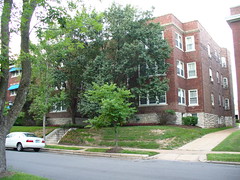
After we were done with work for the day, I took a walk over to the apartment building where most of our lab lived during the hurricane. It was really surreal. On the one hand, all of the Katrina Evacuation, the flooding, the return to New Orleans, etc… feels like a dream. Sometimes I almost believe it didn’t happen, though of course I know it did. And yet, here’s a reminder:

6823 Kingsbury Blvd - Our Home away from Home

We had some good times during our short stay there, and it helped to build camaraderie and cohesion in the lab. We had the whole top floor of that building (two floors) and once even made use of the roof. Shh, don’t tell Quadrangle Housing.

More on open science: PLoS ONE

I wrote last month about my desire for more open access in science, and how PLoS was leading the way. I also said I had some ideas about what more could be done.

It looks like PLoS ONE stole them.

Of course, I jest.

Obviously they’ve been working on this for a while, and I missed it. I only know about it because I’m now subscribed to their RSS feed. This is the framework upon which the future of science publishing will be built. There are many PLoS (Public Library of Science, by the way) journals — PLoS ONE seems to aim to be the open-access equivalent of Science or Nature.

Anyone is allowed to register and comment on or rate articles (with some caveats). They even have guidelines for rating articles. The initial review they mention by an editor and perhaps a few reviewers keeps out the quacks, but anyone is allowed to point out weaknesses in the article or make suggestions on it. It reverses my original idea a bit, in which the article would not be “published” until it had passed a vote by public reviewers, but it is perhaps a more functional model.

I am planning to register and see about reviewing and commenting on some articles. Are you?

Diagnosis Wenckebach

Okay, so this made the rounds a little while ago, but I’m posting it nonetheless:

Amanda and I had a little bit of a communication difficulty when discussing this video. Apparently in medical terminology, at least for a 4th-year medical student, “Wenckebach” is only heard when referring to intermittent AV-nodal block (as deftly illustrated in the video). On the other hand, experimental electrophysiologists frequently refer to any situation in which one area is activating more rapidly than an adjacent area because of differences in refractory period “Wenckebach rhythm”, or if they’re more precise, “Wenckebach-like rhythm”. Many synonyms are also used.

Below is a video of electrical activation during ischemia, illustrating a Wenckebach-like rhythm.

As you can see in the video, the center of the model is only activated every other beat, while the periphery is activated every beat. This is typically referred to as “2:1 capture” or “Wenckebach-like rhythm”, even though the actual phenomenon has very little to do with a true Wenckebach rhythm, in which the atria are activated more rapidly than the ventricles, with block occurring intermittently at the AV-node. The “Diagnosis Wenckebach” has some cute diagrams and even stadium-wave-like demonstrations of this phenomenon.

Long Q-T Syndrome

Today, while looking for blog posts about ICDs and cardiac arrhythmias, I came across the Long Q-T Syndrome Blog. The first story I found was by a woman with long Q-T syndrome who had recently had her ICD replaced in Norway.

Once I checked out the blog itself, I found that it’s done by a group of contributors, and is associated with a long Q-T site, QTsyndrome.ch, which has apparently been around for 10 years! In the past I’ve focused rather narrowly on computational cardiac electrophysiology, and had been frustrated by the lack of other bloggers covering the same field. However, I’m starting to find that while cardiologists may not be predisposed to blogging, patients are! Below are two YouTube videos, found via the Long Q-T Syndrome Blog, on long Q-T syndrome. Neither of them does a great job of explaining what the syndrome is caused by at a low level, but there’s a good FAQ on it here, as well as a short history of the disease and some background on the genetics of inherited long Q-T syndrome.

Those videos also don’t do a good job of explaining what the Q-T interval is. In a somewhat-simplified explanation, it’s the time between when your heart’s ventricles (the major pumps) start contracting, and when they stop contracting and rest. A more detailed explanation of the different parts of an ECG trace, including the Q-T interval, is available on the ECG Learning Center.

And now, the videos:

Anatomical Reentry – 1/10th Speed

This video shows an example of simulated anatomical reentry. The view is of a sheet of simulated tissue with a hole in the middle. A wave of electrical activity continuously circles the hole. This is a stable phenomenon, particularly in this simulation. It should go on indefinitely, as long as I let the simulation keep going. Even in experimental set-ups (such as animal models) these can run for more than an hour.

In the video, black is tissue at rest, white/yellow tissue is activated, and red marks activation (at the front) and re-polarization (at the tail of the wave). If this happened around an obstacle in your heart, it would result in atrial flutter (if it occurred in your atria) or ventricular tachycardia (if it occurred in the ventricles). I’ll do a more extensive post in my “CEP Basics” pages later, but for now, here’s the video. The quality was great until YouTube crappified it.